
Hub · Peptide Science
What is a signal peptide, and how does it direct a protein?
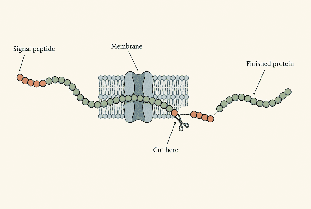
A signal peptide is a short stretch of amino acids, usually at the start of a newly made protein. It works like an address label. It sends the growing chain into the cell's protein delivery system. In mammal cells, sorting machinery brings the chain to the endoplasmic reticulum, a network of membranes inside the cell. There, a channel lets the protein in, and the label is often cut off.
Your saliva holds proteins that were built inside cells and then shipped out of them. Building the right chain is only half the job; the protein also has to reach the right place. Cells read short address labels while proteins are still being made, a bit like a mail room inside every cell. This page in the peptide science hub follows that hidden sorting work.
Key facts
| Subject | Protein targeting signal |
|---|---|
| Building units | Amino acids |
| Typical position | N-terminus: the beginning of the protein chain |
| Main pathway covered | Entry into the mammalian endoplasmic reticulum |
| Recognition machinery | Signal recognition particle (SRP) |
| Membrane channel | Sec61 |
| Sequence length | Variable; no single universal length (von Heijne, 1985) |
What is a signal peptide in simple terms?
A signal peptide is an address written into the protein chain itself. It is not a separate message sent from one cell to another. The address points the protein towards machinery that moves it across a membrane, the thin wall around a cell compartment. In classic experiments from 1975, Blobel and Dobberstein saw an antibody light chain enter membrane vesicles, which are small sealed sacs. Globin, a protein made to stay inside the cell, did not go in. Those results backed the idea that the chain carries its own sorting information. So the term names a part of a protein with a delivery job, not a separate chemical ingredient.
Peptides and proteins are built from the same kind of chain. The peptide and protein comparison explains where one name ends and the other begins. A short stretch can have a peptide name even while it is part of a bigger protein.
What does a signal peptide sequence look like?
A signal peptide sequence is the order of amino acids in the address segment, and different proteins carry different sequences. In 1985, von Heijne compared many of these sequences. He found three regions: a charged start, a middle that avoids water, and a more water-friendly end. Scientists call the water-avoiding part hydrophobic and the water-friendly part polar. The study also found that length and make-up vary a lot. Think of addresses in different handwriting: the layout is similar, but the letters differ. The shared chemical pattern matters more than one fixed string of letters. No single sequence is shared by every signal peptide.
Amino acids are the chain's building blocks, and peptide bonds join them. The N-terminus is the start of the chain. Sequence letters list the amino acids in order. They are not a picture of the finished, folded protein.
How does a cell recognise the signal peptide?
A helper called the signal recognition particle, or SRP, spots the address while the protein is still being built. Building happens on a ribosome, the cell's protein-making machine. In 1981, Walter, Ibrahimi and Blobel showed that SRP binds to ribosomes making a protein meant for release from the cell. They ran this test in a cell-free system, outside living cells. That choosiness separated chains headed for transport from chains made to stay inside the cell. Later work showed that SRP contains both protein and RNA, a nucleic acid that SRP needs for its structure and its job. Put simply, SRP reads the address; it is not the address.
How does the signal peptide direct a protein to the endoplasmic reticulum?
The signal peptide sends a protein to the endoplasmic reticulum, or ER, by engaging sorting machinery. That machinery brings the ribosome and its growing chain to the ER membrane. The ER is a network inside the cell where many proteins that will leave the cell, or sit in its membranes, enter their processing pathways. In rebuilt test systems, adding SRP brought back the choosy binding of these ribosomes to membranes taken from the ER. The binding depended on the transport machinery. So entry is a selective, organised step, like a parcel passing a sorting desk. The protein does not simply slip through the membrane on its own.
Protein targeting is one specific meaning of signalling. What peptides do covers the wider range of peptide roles, including messages that travel between cells. Those roles should not be treated as the same thing.
How does the protein cross the ER membrane?
The growing protein crosses the ER membrane through Sec61, a channel made of protein. The channel opens in response to the signal sequence. In 2016, Voorhees and Hegde captured an active channel using cryo-electron microscopy, a way of imaging frozen molecular samples. The signal sat in a side opening called the lateral gate. Its arrival there was linked to opening a passage across the membrane. This gives a physical picture of how reading the address leads to entry. The signal engages the gate, a bit like a key card at a door, while the rest of the chain passes through the channel into the ER.
Is the signal peptide removed from the finished protein?
A cleavable signal peptide is cut off as the protein enters the ER. Cleavage means cutting the chain at one exact spot. In their rebuilt system, Blobel and Dobberstein found that antibody light chains became shorter as they moved into membrane vesicles. Under those test conditions, the cut happened while the chain was still being made. This explains why the starting protein sequence can include an address piece that the finished protein lacks. It is like peeling a shipping label off a parcel once it arrives. Delivery and trimming are linked events, but the label and the finished protein have different jobs.
Evidence limits
Is a protein signal peptide the same as a cosmetic signal peptide?
No. A protein address signal and a peptide called a signal in cosmetic writing are different ideas. Evidence about one does not establish the role of the other. The address described here is used during protein transport. A different research example is KTTKS, a fragment of a collagen precursor. Collagen is the structural protein in connective tissue, and a precursor is the larger starting molecule. In 1993, Katayama and colleagues studied that fragment in cells grown in a dish. It came from a region cut away from the precursor outside cells. That study did not show KTTKS acting as an ER address. Cell results alone do not establish effects on human skin.
A peptide's name alone does not tell you its mechanism or its evidence. The introduction to peptides explains the broad chemical family. For an unfamiliar sequence, its exact destination and job are not established just because a description uses the word signal.
Comparison
What each named thing does in the cited transport and cell studies. Sharing the word signal does not mean sharing a mechanism.
| Name | Its job | Key study |
|---|---|---|
| Signal peptide | Address label built into a protein chain | Blobel and Dobberstein, 1975 |
| SRP | Reader that spots the address on a growing chain | Walter and colleagues, 1981; Walter and Blobel, 1982 |
| Sec61 | Channel through the ER membrane | Voorhees and Hegde, 2016 |
| KTTKS | Collagen precursor fragment studied in cells | Katayama and colleagues, 1993 |
FAQ
Is a signal peptide the same as a signal sequence?
Signal sequence often names the stretch of amino acids that acts as a signal peptide. In wider writing, targeting signals can mean other sorting information too, so the destination and transport system need naming. This page covers entry into the ER.
Does every signal peptide have the same amino acid sequence?
No. Signal sequences vary in length and chemical make-up. Von Heijne's 1985 analysis found shared regional patterns alongside a lot of variation. A shared transport job does not need an identical chain of amino acids.
Is the signal recognition particle itself a signal peptide?
No. SRP is a combination of protein and RNA that reads transport information on a growing chain. The signal peptide is part of that chain. Walter and Blobel showed in 1982 that RNA is an essential part of SRP.
Does recognising a signal peptide prove where a protein ends up?
No. Recognition and final location are different observations. The studies cited here tested binding, membrane entry or a channel structure. They explain parts of transport, but they do not establish the final destination of every protein described as having a signal.
More in this hub
- Which peptides are approved in Australia and by the FDA?
- What are peptides? Meaning, medicines, safety and legal status in Australia
- What is a peptide bond? Peptide structure, chemistry and synthetic peptides explained
- Citrullinated peptide antibodies: what anti-CCP is, why the test peptide is cyclic and how it is pronounced
- Peptide vs protein: what is the difference? Peptides compared with proteins, hormones, collagen and approved medicines
- What do peptides do? What published studies found
- Peptide side effects: what studies and regulators have recorded
- Do peptides show up on drug tests? How peptides are tested and labelled
- What have collagen peptide studies measured about muscle?
- What is carnosine, and what do studies measure?
- What is CGRP, and how does its signalling work?
- What is icotrokinra, and what do receptor studies measure?
- What is irisin, and how is it studied?
- What is lunasin, and what have studies measured?
- What is pramlintide, and how does amylin signalling work?
- What is peptide YY, and what do studies measure?
- What does TAT-DATNT refer to in peptide research?
Citations
7 sourcesBlobel G, Dobberstein B Journal of Cell Biology. 1975;67(3):852–862.
doi:10.1083/jcb.67.3.852von Heijne G Journal of Molecular Biology. 1985;184(1):99–105.
doi:10.1016/0022-2836(85)90046-4Walter P, Ibrahimi I, Blobel G Journal of Cell Biology. 1981;91(2):545–550.
doi:10.1083/jcb.91.2.545Walter P, Blobel G Journal of Cell Biology. 1981;91(2):551–556.
doi:10.1083/jcb.91.2.551Walter P, Blobel G Nature. 1982;299:691–698.
doi:10.1038/299691a0Voorhees RM, Hegde RS Science. 2016;351(6268):88–91.
doi:10.1126/science.aad4992Katayama K, Armendariz-Borunda J, Raghow R, Kang AH, Seyer JM Journal of Biological Chemistry. 1993;268(14):9941–9944.
doi:10.1016/S0021-9258(18)82153-6
Form Laboratories supplies research-use-only reference material. This article is an educational summary of published third-party literature. It is not medical advice, not a use instruction, and research material is not for human or animal use.