
Hub · Peptide Science
What is CGRP, and how does its signalling work?
CGRP, short for calcitonin gene-related peptide, is a chemical messenger made by nerve cells. It binds a receptor on the cell surface built from two proteins, CLR and RAMP1. Receptor studies link that binding to signals inside the cell, including one called cAMP. Separate tissue studies measure responses such as blood vessels widening. These findings describe a body system; they do not establish personal health outcomes.
01CGRP identityContents
Nerves talk in two ways. One is the quick electrical pulse most people picture. The other is chemical: nerve cells make messenger molecules that land on receptors on other cells. CGRP is one of those chemical messengers. This page traces its story from gene to cell surface, as part of the peptide science hub.
Key facts
| Abbreviation | CGRP |
|---|---|
| Full name | Calcitonin gene-related peptide |
| Class | Neuropeptide: a peptide messenger made by nerve cells |
| Gene-processing study | Rosenfeld et al., Nature, 1983 |
| Receptor components | CLR and RAMP1 (McLatchie et al., Nature, 1998) |
| Cell signal measured | cAMP accumulation (Muff et al., FEBS Letters, 1998) |
What is the CGRP peptide?
CGRP is a neuropeptide: a short chain of amino acids that nerve cells make and use as a chemical messenger. The full name is calcitonin gene-related peptide. In 1983, Rosenfeld and colleagues described how nerve tissue makes CGRP. They linked a gene's RNA message to the peptide made from that message. RNA is a working copy of a gene's instructions, like a photocopy of one page from a recipe book. The team also mapped which cells and pathways contain CGRP. That map pointed to possible roles worth studying. But where a messenger is found does not, by itself, prove what it does.
Peptide describes the kind of molecule. Neuropeptide adds where the messenger comes from. The peptide definition page explains the building blocks, while the peptide bond explanation describes the chemical link that joins them.
Is CGRP the same as calcitonin?
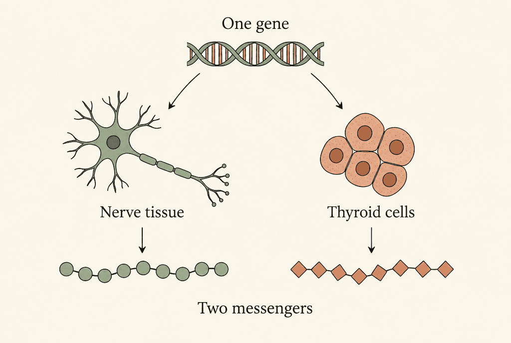
No. CGRP and calcitonin are different messengers, even though the original research traced them to the same gene. Rosenfeld and colleagues found that nerve tissue and thyroid C cells process that gene's RNA in different ways. Nerve tissue made a message for the CGRP precursor. Thyroid C cells made a message for the calcitonin precursor. A precursor is a larger starting molecule that is later shaped into the finished messenger. Think of one recipe book used by two kitchens, each making a different dish. The shared gene explains the name. It does not mean the two finished peptides carry the same message.
Which receptor recognises CGRP?
The CGRP receptor has two parts: CLR, short for calcitonin receptor-like receptor, and RAMP1, short for receptor activity-modifying protein 1. A receptor is a protein system that receives a signal at the cell surface. In 1998, McLatchie and colleagues showed that the partner protein matters. It changes how CLR reaches the cell surface and which peptide the pair recognises. Paired with RAMP1, CLR formed a CGRP receptor. Paired with RAMP2, CLR formed a receptor for a related messenger called adrenomedullin. So the same main protein can receive different messages, depending on which partner it has.
Receptor identity helps explain why one broad label, peptide, covers so many different signals. The overview of peptide functions places receptor signalling alongside the other jobs peptides do in the body.
How does CGRP binding become a signal inside a cell?
When CGRP binds its receptor, a message outside the cell connects to signalling machinery inside it. In 2018, Liang and colleagues imaged CGRP bound to CLR, RAMP1 and Gs, a relay protein inside the cell. They used cryo-electron microscopy, a method that builds a molecular image from frozen samples. The image showed where the peptide touches the receptor complex, with RAMP1 helping to hold the receptor steady. It captures an active molecular arrangement, like a single photo of a handshake. A still image like this is not a film of a nerve signal moving through a living person.
Cell experiments give a separate measure of receptor activity. In 1998, Muff and colleagues recorded a rise in cAMP in rabbit aortic endothelial cells that carried RAMP1. Endothelial cells line blood vessels. cAMP, short for cyclic adenosine monophosphate, is a messenger inside cells. A receptor blocker worked against the response, which supports the receptor link within that cell model.
What do physiological studies of CGRP measure?
Body-tissue studies of CGRP measure things like vessel width, blood flow and changes in how tense a vessel wall is. In 1985, Brain and colleagues reported more local blood flow in rabbit skin. They also saw small arteries widen in hamster tissue, and a piece of rat aorta, the body's main artery, relax outside the body. The study recorded local redness in human skin too. Each result belongs to its own species, tissue and test setup. Together, they support a role for CGRP in blood-vessel signalling. They do not show that all tissues respond alike, or that vessel widening predicts a health outcome.
The measurement matters as much as the molecule's name. Blood flow, vessel tension and cAMP each answer a different question. Peptides and proteins compared gives more background on why a molecule's label alone cannot describe a response in the body.
Evidence limits
What can CGRP mechanism studies establish?
CGRP mechanism studies show how one part of the system works, but a receptor result alone does not establish a personal health outcome. A receptor image shows how molecules fit together. A cell experiment measures a response within that cell system. A tissue experiment records what happens in one specific tissue. These questions sit at different levels of biology, like a street map, a single house and one room. Liang and colleagues examined the active human receptor complex in 2018. Brain and colleagues studied blood-vessel responses in 1985. Neither finding, by itself, answers every question about CGRP across the whole human body.
Evidence keeps its meaning when the measurement stays attached to its model. A change in a cell signal is not the same measurement as a change in blood flow. This page names the model beside each finding. It does not treat a supplied research material as equal to the body's own signalling system.
Comparison
Molecules named in the cited gene and receptor studies. The table compares what each one is, not health outcomes.
| Molecule | What it is | Key study |
|---|---|---|
| CGRP | Messenger made in nerve tissue from the shared gene | Rosenfeld et al., 1983 |
| Calcitonin | Separate messenger made in thyroid C cells from the same gene | Rosenfeld et al., 1983 |
| CLR with RAMP1 | Receptor pair that recognises CGRP | McLatchie et al., 1998 |
| CLR with RAMP2 | Receptor pair that recognises adrenomedullin | McLatchie et al., 1998 |
FAQ
What does CGRP stand for?
CGRP stands for calcitonin gene-related peptide. The name reflects the shared gene that Rosenfeld and colleagues described in 1983. It does not mean CGRP is the same as calcitonin.
Is CGRP a peptide or a receptor?
CGRP is the peptide messenger. CLR together with RAMP1 forms the CGRP receptor, which receives the message at the cell surface, as McLatchie and colleagues showed in 1998.
What is cAMP in CGRP research?
cAMP is cyclic adenosine monophosphate, a messenger inside cells. Muff and colleagues measured a CGRP-linked rise in cAMP in rabbit vascular cells carrying RAMP1. That result describes that cell system.
Does a blood-vessel response prove a health outcome?
No. A blood-vessel response shows a measured change in the tissue studied. The vessel and skin findings that Brain and colleagues reported in 1985 do not, on their own, establish a person's health outcome.
More in this hub
- Which peptides are approved in Australia and by the FDA?
- What are peptides? Meaning, medicines, safety and legal status in Australia
- What is a peptide bond? Peptide structure, chemistry and synthetic peptides explained
- Citrullinated peptide antibodies: what anti-CCP is, why the test peptide is cyclic and how it is pronounced
- Peptide vs protein: what is the difference? Peptides compared with proteins, hormones, collagen and approved medicines
- What do peptides do? What published studies found
- Peptide side effects: what studies and regulators have recorded
- Do peptides show up on drug tests? How peptides are tested and labelled
- What have collagen peptide studies measured about muscle?
- What is a signal peptide, and how does it direct a protein?
- What is carnosine, and what do studies measure?
- What is icotrokinra, and what do receptor studies measure?
- What is irisin, and how is it studied?
- What is lunasin, and what have studies measured?
- What is pramlintide, and how does amylin signalling work?
- What is peptide YY, and what do studies measure?
- What does TAT-DATNT refer to in peptide research?
Citations
5 sourcesRosenfeld et al. Nature. 1983;304:129-135.
doi:10.1038/304129a0McLatchie et al. Nature. 1998;393:333-339.
doi:10.1038/30666Liang et al. Nature. 2018;561:492-497.
doi:10.1038/s41586-018-0535-yMuff et al. FEBS Letters. 1998;441(3):366-368.
doi:10.1016/S0014-5793(98)01587-7Brain et al. Nature. 1985;313:54-56.
doi:10.1038/313054a0
Form Laboratories supplies research-use-only reference material. This article is an educational summary of published third-party literature. It is not medical advice, not a use instruction, and research material is not for human or animal use.